Download præsentationen
Præsentation er lastning. Vent venligst

1
Immunsystemet og Biologiske lægemidler
9 maj 2012

3
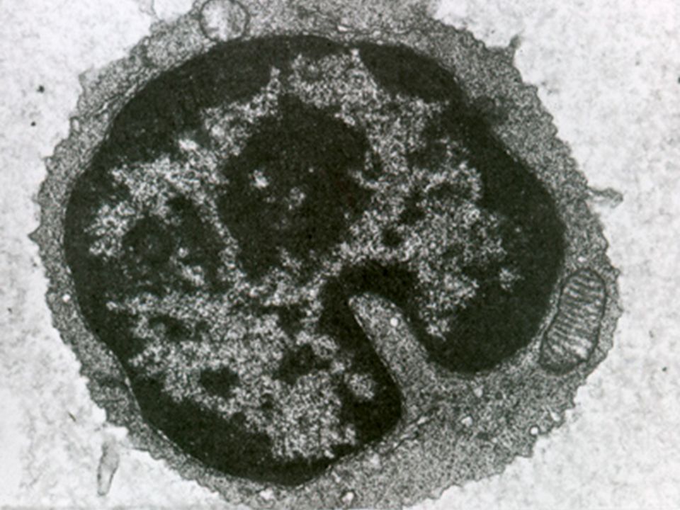
Figur 1. Fagocytose

4
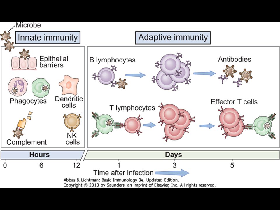
Figur 2. Det adaptive immunsystem, organer

5
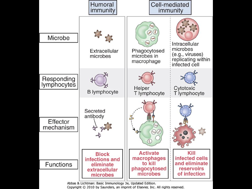
Figur 3. Det adaptive immunsystem, celler

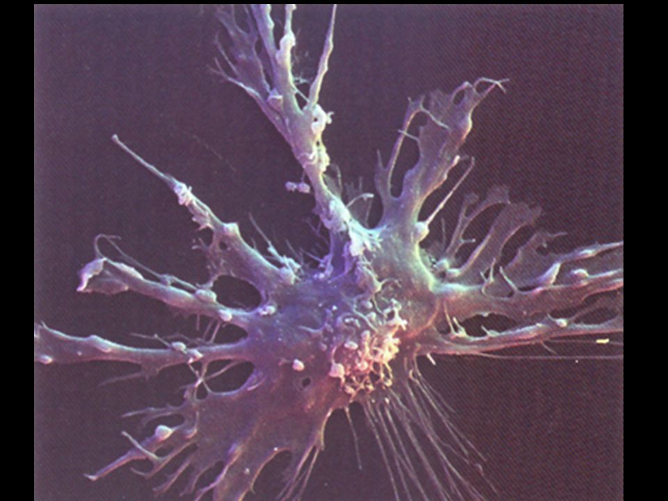
7
Dendrit celle

10
Figur 6. Immunologisk hukommelse

11
Figur 5. Proteinantigen

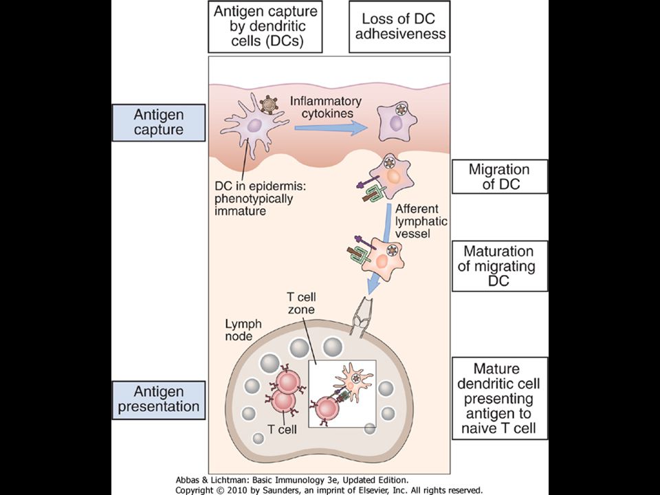
13
Figur 7. Præsentation af antigen for T-lymfocytter

14
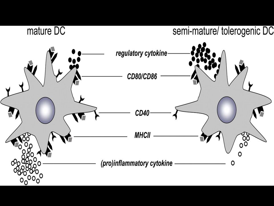
Dendritiske celler i hud

16
Figur 4. Lymfocytternes recirkulation

19
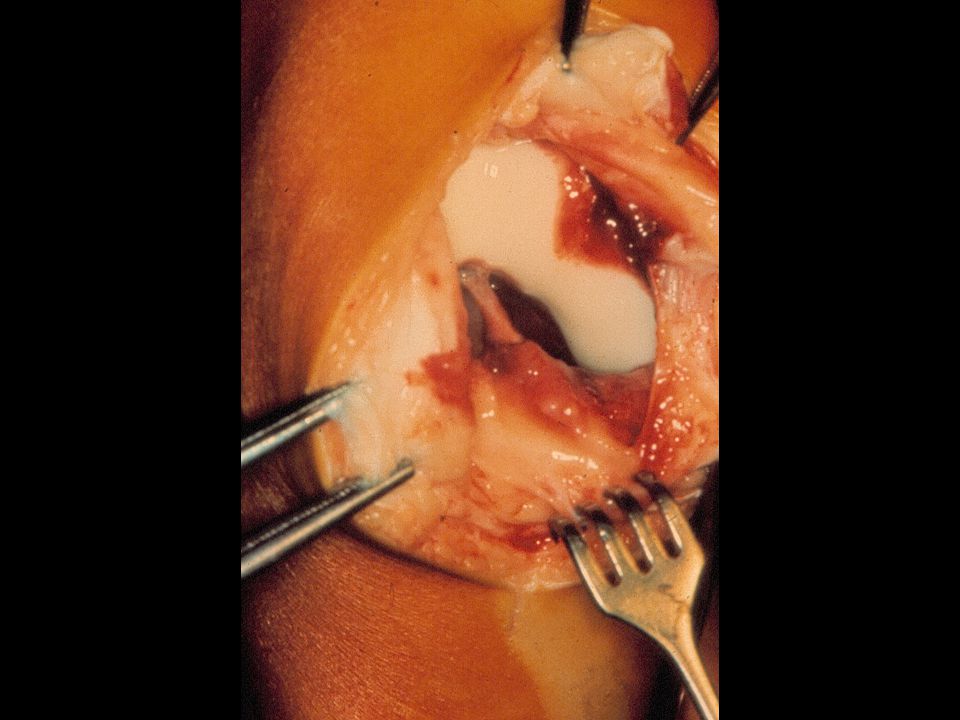
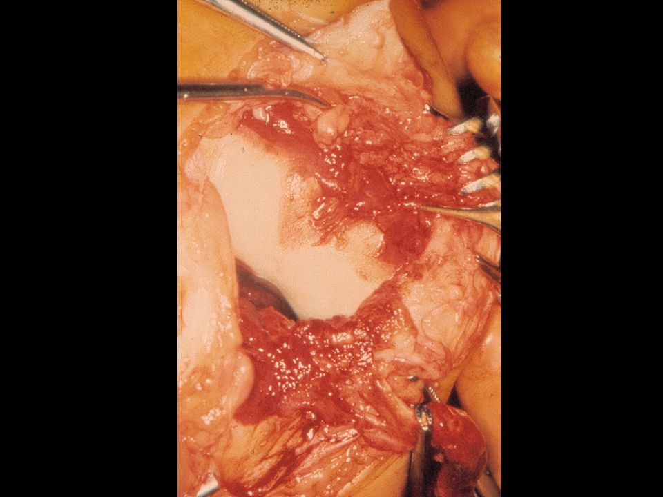
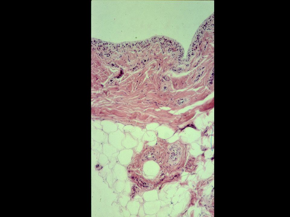
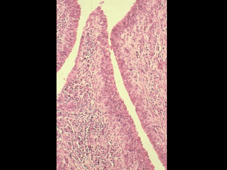
Tidlig RA

24
IL-1b and TNF-a: Proinflammatory Cytokines in the Rheumatoid Joint
High endothelial venule B o n e O s t e o b l a s t s O s t e o c l a s t s Synovial membrane IL-8 C a r t i l a g e PGE2 IL-6 N e u t r o p h i l s Capsule T N F - a I L - 1 Synovial space Interleukin-1 (IL-1) and tumour necrosis factor- (TNF-) have been identified as pivotal proinflammatory cytokines in the pathogenesis of the rheumatoid joint.1,2 In this slide, IL-1 and TNF- are shown in the joint space; however, concentrations of these cytokines are likely to be higher in the tissues.1 Increased concentrations of IL-1 and TNF- are found in the synovial fluid and tissue of patients with rheumatoid arthritis (RA). These cytokines act to stimulate the production of each other, that is, IL-1 stimulates production of TNF- and vice versa.1 Early in the RA disease process, IL-1 and TNF- act synergistically to increase production of matrix metalloproteases, such as collagenase, by chondrocytes. These enzymes degrade components of the cartilage matrix.1 IL-1 also activates osteoclasts in bone.2 IL-1 and TNF- also increase expression of adhesion molecules on the endothelium, contributing to the migration of neutrophils and lymphocytes from the circulation.1 In addition, IL-1 and TNF- stimulate synovial fibroblasts to produce additional proinflammatory mediators, such as IL-8, prostaglandin-E2, and IL-6. These mediators are responsible for the acute and chronic inflammation characteristic of RA.1 1. Dinarello CA. Biologic basis for interleukin-1 in disease. Blood. 1996;87: 2. Gravallese EM, Goldring SR. Cellular mechanisms and the role of cytokines in bone erosions in rheumatoid arthritis. Arthritis Rheum. 2000;43: 3. Dinarello CA, Moldawer LL. Proinflammatory and Anti-inflammatory Cytokines in Rheumatoid Arthritis. A Primer for Clinicians. 3rd ed. Thousand Oaks, Ca: Amgen Inc.; 2001. C h o n d r o c y t e s Pannus Osteoblasts Osteoclasts B o n e PGE2 = prostaglandin-E2 Dinarello C, Moldawer L. Proinflammatory and Anti-inflammatory Cytokines in Rheumatoid Arthritis: A Primer for Clinicians. 3rd ed. Thousand Oaks, Ca, USA: Amgen Inc.; 2001.
 and tumour necrosis factor- (TNF-) have been identified as pivotal proinflammatory cytokines in the pathogenesis of the rheumatoid joint.1,2. In this slide, IL-1 and TNF- are shown in the joint space; however, concentrations of these cytokines are likely to be higher in the tissues.1. Increased concentrations of IL-1 and TNF- are found in the synovial fluid and tissue of patients with rheumatoid arthritis (RA). These cytokines act to stimulate the production of each other, that is, IL-1 stimulates production of TNF- and vice versa.1. Early in the RA disease process, IL-1 and TNF- act synergistically to increase production of matrix metalloproteases, such as collagenase, by chondrocytes. These enzymes degrade components of the cartilage matrix.1. IL-1 also activates osteoclasts in bone.2. IL-1 and TNF- also increase expression of adhesion molecules on the endothelium, contributing to the migration of neutrophils and lymphocytes from the circulation.1. In addition, IL-1 and TNF- stimulate synovial fibroblasts to produce additional proinflammatory mediators, such as IL-8, prostaglandin-E2, and IL-6. These mediators are responsible for the acute and chronic inflammation characteristic of RA Dinarello CA. Biologic basis for interleukin-1 in disease. Blood. 1996;87: Gravallese EM, Goldring SR. Cellular mechanisms and the role of cytokines in bone erosions in rheumatoid arthritis. Arthritis Rheum. 2000;43: Dinarello CA, Moldawer LL. Proinflammatory and Anti-inflammatory Cytokines in Rheumatoid Arthritis. A Primer for Clinicians. 3rd ed. Thousand Oaks, Ca: Amgen Inc.; C. h. o. n. d. r. o. c. y. t. e. s. Pannus. Osteoblasts. Osteoclasts. B. o. n. e. PGE2 = prostaglandin-E2. Dinarello C, Moldawer L. Proinflammatory and Anti-inflammatory Cytokines in Rheumatoid Arthritis: A Primer for Clinicians. 3rd ed. Thousand Oaks, Ca, USA: Amgen Inc.;")
25
TNF alfa i RA Produktion af kemokiner og adhæsionsmolekyler
Produktion af andre proinflammatoriske cytokiner Produktion af metalloproteinaser og PGE2 Nedbrydning af brusk og knogle

26
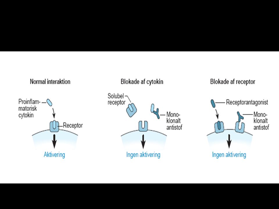
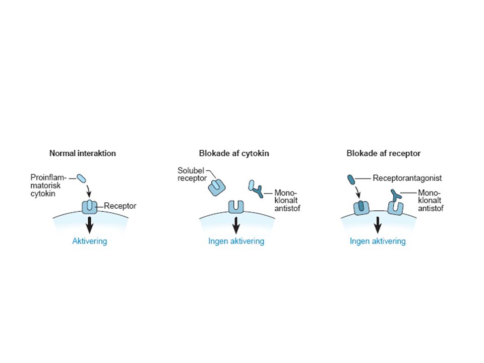
Figur 13. Basisenheden i immunglobulin molekylet

28
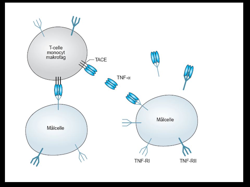
Although the etiology of rheumatoid arthritis remains unknown, there is good evidence to suggest that the host immune response may play a significant role in the perpetuation of chronic inflammation. TNFa is produced predominantly in monocytes, macrophages and activated T cells and has been found to participate in chronic inflammatory disease processes, particularly those depending upon collaboration between T cells and macrophages. TNFa is expressed as a 26 kD membrane protein which upon proteolysis is released as a 157 amino acid, 17 kD monomeric protein. These monomers self-associate into a homotrimer which is the active form of soluble TNFa. Both soluble TNFa and membrane-bound TNFa have biologic activity. Two different receptors for TNFa exist: TNF-R p55 and TNF-R p75. Membrane bound receptors for TNFa have been identified on a wide variety of tissue and cell types. TNFa trimer can bind as many as three TNF receptors and this cross-linking of receptors initiates signal transduction within the target cell. Binding of TNFa to TNF-R p55 induces cytotoxicity, fibroblast proliferation, synthesis of prostaglandins, up-regulation of adhesion molecules, NFB activation, etc. The role of TNF-R p75 is less well defined. It appears to concentrate soluble TNFa at the cell surface for transfer to TNF-R p55 and may prefer the transmembrane form of TNFa for signaling.

29
Antibody Neutralization of TNF
Infliximab binds to released or soluble TNFa, membrane bound TNFa, as well as TNFa bound to target cells. It has been shown, in vitro, that the binding of infliximab to the membrane bound form either alters the function of the TNFa producing cell or results in lysis through complement activation or antibody-dependent cellular toxicity. Also, infliximab prevents the interaction of TNFa with its cellular receptors, TNF-R p55 and TNF-R p75. In in vitro tests, infliximab inhibits the binding to both receptors, suggesting that infliximab can inhibit TNFa-mediated signaling through either receptor in vivo. Neutralization of TNFa action has been suggested as a mode of therapeutic intervention in a variety of human diseases as a result of emerging data implicating TNFa as an important pathophysiological regulator. This inhibition of TNF activity does not appear to result in generalized immunosuppression.

32
Etanercept ENBREL® (etanercept) is a recombinant soluble TNF-receptor formed by the fusion of two human TNF-receptors and the Fc portion of human IgG1. The diagram illustrates the dimeric structure of ENBREL, engineered to bind TNF with high affinity.1 Reference: 1. ENBREL Prescribing Information, Immunex Corporation, Seattle, Wash.
 is a recombinant soluble TNF-receptor formed by the fusion of two human TNF-receptors and the Fc portion of human IgG1. The diagram illustrates the dimeric structure of ENBREL, engineered to bind TNF with high affinity.1. Reference: 1. ENBREL Prescribing Information, Immunex Corporation, Seattle, Wash.")
33
Receptor Fusion proteins: Mechanism of action
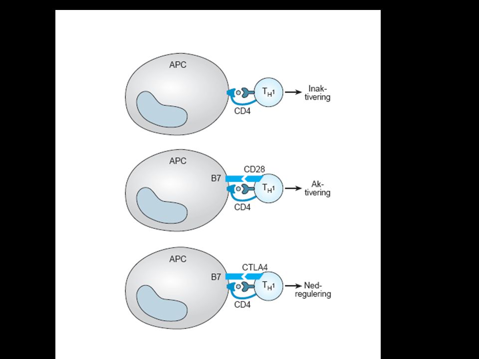
Hendricks

34
* The biologic role of a Ab is to capture and retain
The role of receptors is to capture and release, thereby triggering a signal

35
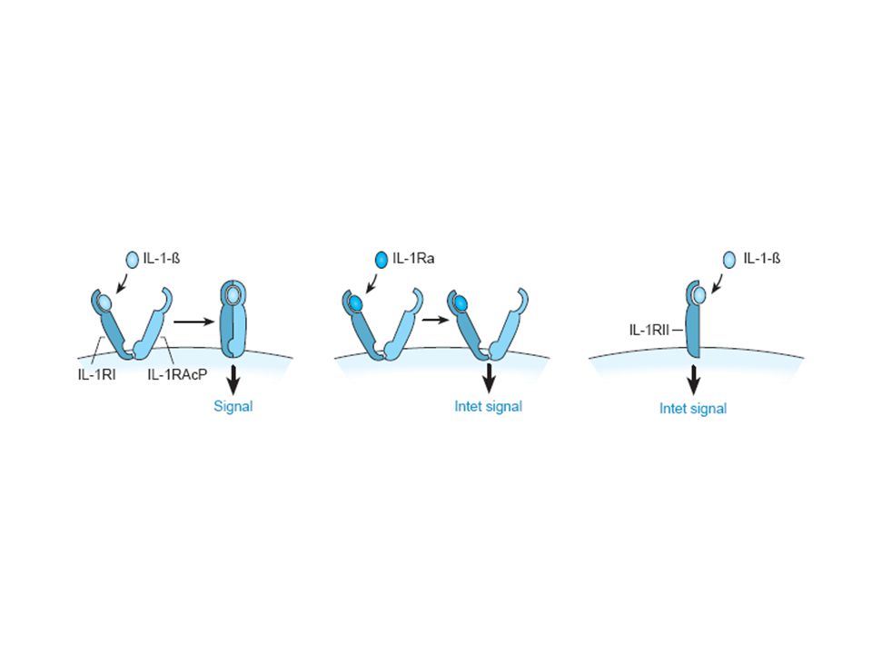
anakinra

38
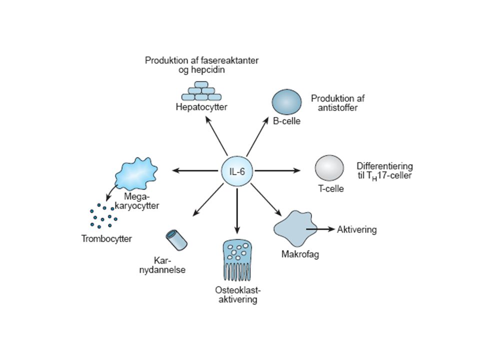
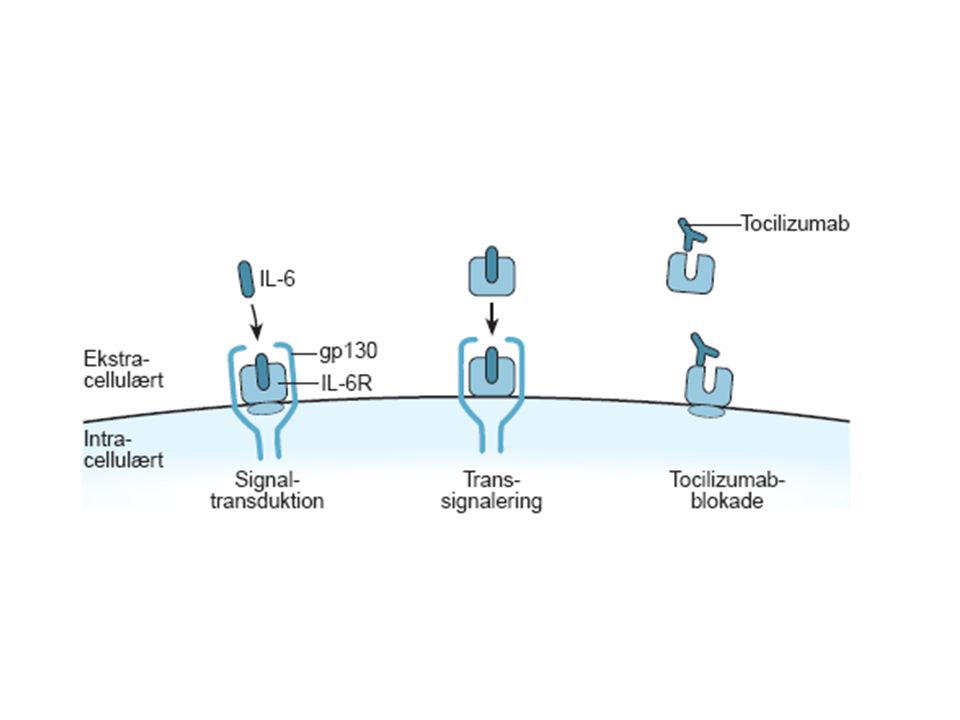
tocilizumab

42
Cytokine Signaling Pathways Involved in Rheumatoid Arthritis
TNF-a IL-1 IL-6 IFN-g IL-12 IL-4 IL-10 Macrophage RF Plasma cell B cell Interferon-g Th0 Th2 Synovium OPGL CD4 + T cell CD69 CD11 Osteoclast Fibroblast Chondrocyte Production of metalloproteinases and other effector molecules Migration of polymorphonuclear cells Erosion of bone and cartilage TNF- has a primary role in the pathogenesis of rheumatoid arthritis. TNF- and its downstream cytokines are potent stimulators of mesenchymal cells, such as synovial fibroblasts, osteoclasts and chondrocytes that release tissue-destroying matrix metalloproteinases which ultimately lead to the erosion of bone and cartilage. Choy EHS, Panayi GS. Cytokine pathways and joint inflammation in rheumatoid arthritis. N Engl J Med. 2001;344: Choy EHS, Panayi GS. N Engl J Med. 2001;344: © 2001 Massachusetts Medical Society. All rights reserved.

43
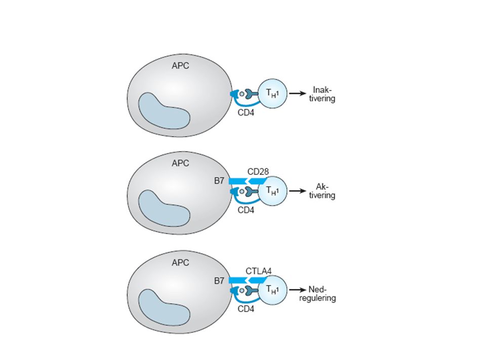
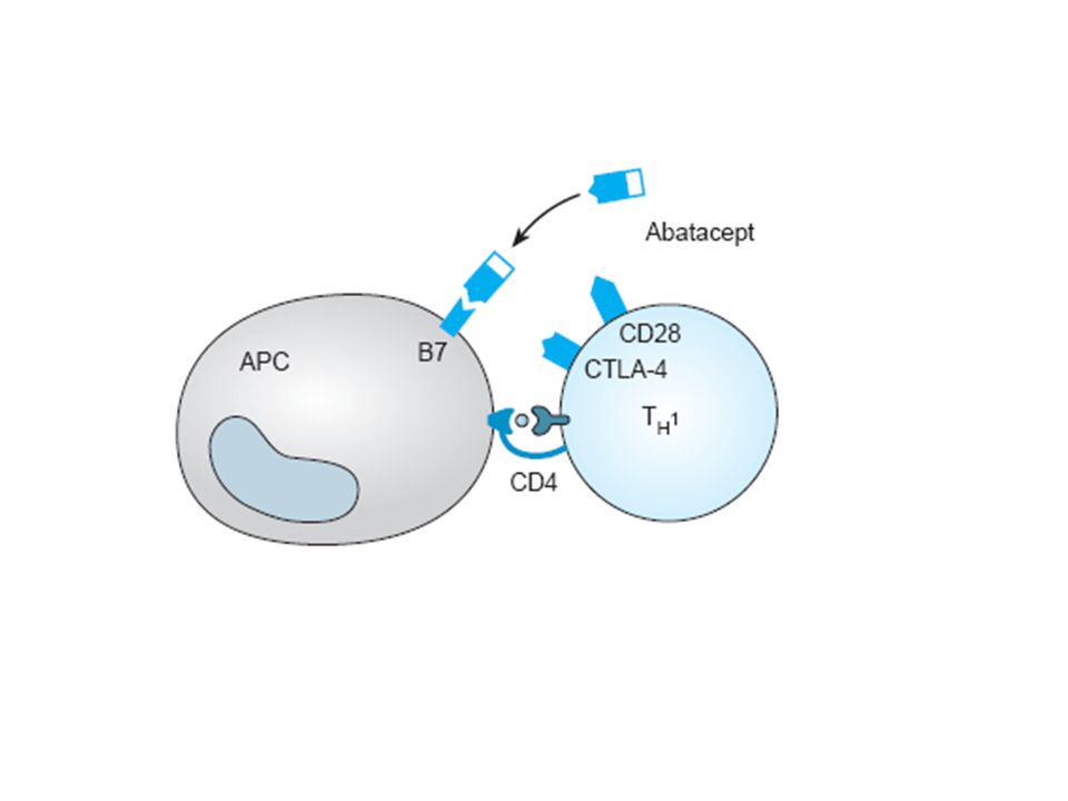
abatacept

45
Abatacept (CTLA-4Ig) is a Recombinant Human Fusion Protein Comprised of CTLA-4 and a Modified Fc Domain of IgG-1 CTLA-4 IgG-1 Abatacept (CTLA-4Ig) Extracellular Cell membrane Intracellular This slide shows the structure of abatacept, which is a fully human fusion protein comprised of 2 domains. Abatacept : fully human fusion protein is less immunogenetic than chimeric human-murine monoclonal antibody. The first domain is the extracellular domain of human CTLA-4 which binds to CD80/86. Because only the extracellular domain is used, binding to CD80/86 can occur, but no negative signaling occurs since the intracellular domain is missing. As is done with many fusion proteins where a transmembraneous domain is removed, an Fc domain is used to stabilize the protein and optimize recombinant production and purification. The Fc domain of abatacept is derived from human IgG1. It is important to note that this Fc domain is inactive as is described on the next slide. Fc Modified Linsley PS et al. J Exp Med. 1991; 174: 561–9. Ig=immunoglobulin.
 Extracellular. Cell membrane. Intracellular. This slide shows the structure of abatacept, which is a fully human fusion protein comprised of 2 domains. Abatacept : fully human fusion protein is less immunogenetic than chimeric human-murine monoclonal antibody. The first domain is the extracellular domain of human CTLA-4 which binds to CD80/86. Because only the extracellular domain is used, binding to CD80/86 can occur, but no negative signaling occurs since the intracellular domain is missing. As is done with many fusion proteins where a transmembraneous domain is removed, an Fc domain is used to stabilize the protein and optimize recombinant production and purification. The Fc domain of abatacept is derived from human IgG1. It is important to note that this Fc domain is inactive as is described on the next slide. Fc. Modified. Linsley PS et al. J Exp Med. 1991; 174: 561–9. Ig=immunoglobulin.")
47
rituximab

49
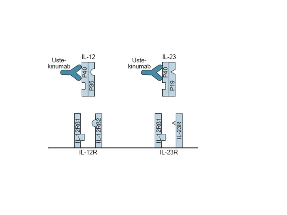
Fuldt humant antistof mod p40 subunit, som er tilstede i både
Ustekinumab Fuldt humant antistof mod p40 subunit, som er tilstede i både IL-12 og IL-23

50
EPIDERMIS Th1 Th17 Naive T-celler Umodne Langerhansceller Migrerende
Il-6, IL-17, IL-21, IL-22 og TNF-α IL-12 INF-γ INF-γ TNF-α Th17 IL-1, IL-6 IL-21, IL-23 og TGF-β CD11c+ dendritiske celler og plasmocytoide dendritiske celler Naive T-celler Blodkar

Lignende præsentationer


 Projektledelse og Produktion af Digitalt Indhold (DPI) Projektledelse IT-projektledelse (ITP) Projektledelse og.>")


